Protein
Synthesis
Transcription & Translation |
The
most important facet of the function of living cells is the
synthesis of proteins. Because proteins carry out
multiple tasks in the body, the mechanism to synthesize them
is highly intricate.
Despite the overall complexity of this process, it occurs
with remarkable accuracy. The rate of error is roughly one
in every 10,000 amino acids. Using the processes of transcription
and translation, the body makes an amazing number and variety
of proteins.
The
transcription and translation processes provide
the correct primary structure of the protein |
The
protein must fold to obtain the correct secondary
and tertiary structures. Protein folding remains an active
research area.
There are several stages involved in the synthesis process,
including transcription and translation.
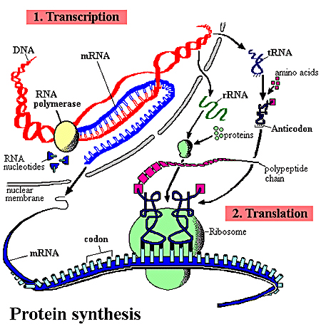
The illustration above (Protein Synthesis) shows the process
whereby DNA encodes for the production of amino acids and
proteins.
This process can be divided into two parts:
1. Transcription
Before the synthesis of a protein begins, the corresponding
RNA molecule is produced by RNA transcription. One strand
of the DNA double helix is used as a template by the RNA polymerase
to synthesize a messenger RNA (mRNA). This mRNA migrates from
the nucleus to the cytoplasm. During this step, mRNA goes
through different types of maturation including one called
splicing when the non-coding sequences are eliminated. The
coding mRNA sequence can be described as a unit of three nucleotides
called a codon.
The primary role of deoxyribonucleic acid (DNA) is to direct
the synthesis of proteins. DNA, however, is located in the
nucleus of the cell, and protein synthesis occurs in cellular
structures called ribosomes, found out-side the nucleus. The
process by which genetic information is transferred from the
nucleus to the ribosomes is called transcription. During transcription,
a strand of ribonucleic acid (RNA) is synthesized. This messenger
RNA (mRNA) is complementary to the portion of DNA that directed
it: as it has a complementary nucleotide at each point in
the chain.
A specialized protein called an enzyme controls when transcription
occurs. The enzyme called RNA polymerase is present in all
cells; eukaryotic cells have three types of this enzyme. DNA
has a section called the promoter region that identifies the
sites where transcription starts and must be recognized by
one subunit of the RNA polymerase called the sigma (s) factor.
Recognition between the promoter and the s-factor helps to
regulate how often a particular gene is transcribed. Once
bound, the polymerase initiates the construction of mRNA (or
other RNA molecules).
Initiation of the synthesis of a new RNA molecule does not
always lead to a complete synthesis. After roughly ten nucleotides
have been strung together, the continued addition of complementary
base pairs takes place more readily in a process called elongation.
The speed of addition of new nucleotides is remarkablebetween
twenty and fifty nucleotides per second can be added at body
temperature.
Eventually the elongation process must stop. There are certain
sequences of nucleotides that stop elongation, a process called
termination. Often, termination occurs when the newly formed
section of RNA loops back on itself in a tight formation called
a hairpin. Once the hairpin structure has formed, the last
component is then a string of uracil residues.
After transcription has taken place, the mRNA produced is
not necessarily ready to direct the subsequent protein synthesis.
Depending on the type of cell, segments of nucleotides may
be removed or appended before the actual synthesis process
takes place. This type of post-transcriptional processing
often occurs in human cells.
2. Translation
The ribosome binds to the mRNA at the start codon (AUG) that
is recognized only by the initiator tRNA. The ribosome proceeds
to the elongation phase of protein synthesis. During this
stage, complexes, composed of an amino acid linked to tRNA,
sequentially bind to the appropriate codon in mRNA by forming
complementary base pairs with the tRNA anticodon. The ribosome
moves from codon to codon along the mRNA. Amino acids are
added one by one, translated into polypeptidic sequences dictated
by DNA and represented by mRNA. At the end, a release factor
binds to the stop codon, terminating translation and releasing
the complete polypeptide from the ribosome. One specific amino
acid can correspond to more than one codon. The genetic code
is said to be degenerate.
Once the mRNA has been synthesized, and perhaps modified,
the next step of protein synthesis, translation, takes place.
For this stage, additional forms of RNA are needed.
Transfer RNA (tRNA) plays the role of carrying an amino acid
to the synthesis site at the ribosome. tRNA molecules are
relatively small, with around seventy-five nucleotides in
a single strand. They form several loops, one of which is
an anti-codon, a three-residue series that is complementary
to the codon present in the mRNA.
The opposite end of the tRNA is where an amino acid is bound.
The correct binding of an amino acid to a specific tRNA is
every bit as important as the anti-codon in ensuring that
the correct amino acid is incorporated in the polypeptide
that is synthesized.
There are different tRNA molecules for each of the twenty
amino acids that are present in living systems; some amino
acids have more than one tRNA that carry them to the synthesis
site.
When translation begins, mRNA forms a complex with a ribosome
to form an assembly site. This complex requires the assistance
of proteins called initiation factors, so the existence of
an mRNA does not mean that a protein will always be synthesized.
The first tRNA that takes part in the initiation always carries
the same amino acid, methionine. When the protein is completely
synthesized, this initial methionine is often removed.
With the initial methionine in place, another tRNA with its
amino acid joins the assembly site as dictated by the codon
on the mRNA. With two amino acids present, a peptide bond
can be formed and the polypeptide can begin forming.
The new amino acid is added to the carbon end of the polypeptide
(the C-terminus) with the peptide bond forming between the
C-O of the polypeptide and the amine of the new amino acid.
This structural specificity is enforced by the nature of the
binding between the amino acid and the tRNA. The portion of
the amino acid that is unbound in the tRNA complex is the
amine.
Elongation
ultimately requires the repetition of several steps:
|
| (1)
|
The
tRNAamino acid complexes must be made. |
| (2) |
This
complex must bind to the mRNA-ribosome assembly site.
The correct amino acid is assured by the matching of
the anti-codon on the tRNA to the codon on the mRNA. |
| (3) |
A peptide bond is formed between the new amino acid
and the growing polypeptide chain. |
| (4) |
The amino acid is cleaved from the tRNA, which can be
cycled back to form another complex with an amino acid
for a later synthesis. |
| (5)
|
The
growing polypeptide forms a fiber-like tendril. |
| (6) |
The ribosome essentially moves along the mRNA, reopening
the initiation site for additional protein synthesis.
In this way, proteins are synthesized by several ribosomes
acting on the same mRNA molecule. |
The structure of the ribosome plays an important role in this
elongation process. There must be two sites available for
synthesis to occur. One site, called the P site (for peptide),
is where the growing (or nascent) polypeptide is located.
Adjacent to this location is another site where the tRNA with
its new amino acid can bind.
This site is called the A site (for the amino acid that is
delivered there along with the tRNA).
As was the case in the elongation of mRNA noted earlier, somehow
the emerging polypeptide must stop adding amino acids. The
termination is actually part of the coding present in the
codons. Three specific codons are known as stop codes, and
when they are present in mRNA, the elongation is stopped.
|